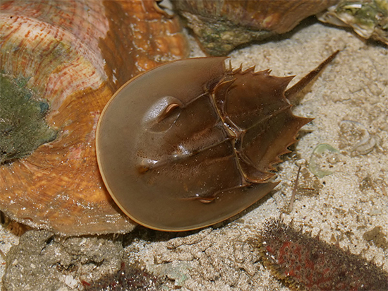
First, horseshoe crabs aren’t crabs at all. Rather, they are arachnids, the same class as spiders and scorpions. What’s more, they have ten eyes! The two lateral eyes are compound eyes, meaning they consist of about 1,000 units, each consisting of a cornea, lens, and cones and rods. (Sound familiar?) These eyes are sensitive to polarized light and can magnify sunlight ten times and are a million times more sensitive to light at night than during the day. Two simple eyes that can sense ultraviolet light from the moon are forward and central to the lateral eyes. Behind the lateral eyes are two primitive eyes and three more along a ridge on the underside of the shell. (Are you counting?) Photoreceptors on the telson (tail) make up the last eye. These photoreceptors are thought to help the horseshoe crab’s brain synchronize to light and dark cycles. With all those eyes and cones and rods the largest of any known animal, 100 times the size of those in humans, their vision is quite good.
 |
For more than fifty years, researchers have studied the horseshoe crab’s optic nerves, some of which are sensitive to light at 535 nanometers (green), and others to 380 nm (HEV). With three different types of eyes, large retinal neurons and well-defined visual behavior, the visual system of horseshoe crabs offers a wealth of information. What’s more, the structure and function of their eyes are regulated on a daily basis by a circadian clock in the animal’s brain. The eye-brain connection of horseshoe crabs is easily available to study and its unique functions have lent a great deal to the study of human vision.
Learn more about human eye development and function with our CE, Check Your Eye Development IQ, at 2020mag.com/ce.