Despite your best efforts intraoperative complications can derail even an apparently routine cataract extraction: The capsule can tear; vitreous can come forward; or an iris can be flaccid and be accompanied by a small pupil. However, surgeons say that if you're prepared, both with tools on hand and techniques in mind, you can manage most of these complications effectively. In this article, a group of accomplished surgeons share their advice on managing several of the complications you're likely to run into in any given month of surgeries.
Anterior Capsular Tears
Surgeons say anterior capsular tears can be managed adequately, but the threat of a posterior tear must always be taken into account.
"Frequently, anterior capsular tears aren't noticed until the case is at the point where the anterior capsule's readily visible," says
"Anterior capsule tears occur during the surgery when there's not a continuous capsulorhexis, which leaves a little nick that can extend," Dr. Fishkind explains. "They can occur during the surgery due to lifting the nucleus with a small capsulorhexis. They can also occur because the anterior capsule is emulsified while the surgeon is emulsifying sub-anterior capsular material, or from positive pressure from the posterior when there's any weakness in the anterior capsule."
Surgeons say that, when you recognize an anterior capsular tear, the key is to prevent it from extending through the equator and into the posterior capsule. "Always maintain a deep anterior chamber," says Dr. Fishkind. "This means that if there's a phaco tip in the eye and you recognize the anterior capsular rent, then, before removing the tip—which would allow shallowing of the chamber and possible extension of the rent—fill the anterior chamber with an occlusive viscosurgical device, either a cohesive or dispersive, and fill it such that when the phaco probe is removed the chamber won't shallow. Then, if there's residual cortex to be removed, it's better to remove it with a manual technique rather than by inserting a large instrument. This is because, again, any time you put an I/A tip in the eye there are going to be fluid fluctuations that allow the chamber to deepen and shallow, creating the type of situation that can allow the anterior capsular tear to extend."

Surgeons say the implantation of the intraocular lens is usually not a serious concern with anterior tears. "I'll still put the lens in the bag," says Dr. Grayson. "I use a one-piece Alcon SN60, but any intracapsular lens is fine. I'll try to orient the haptics in the area that's still very well contained on the anterior capsular rim." Dr. Fishkind also thinks a capsular bag placement is feasible. "If I'm concerned that the capsule is less intact, then I'll put a three-piece lens into the capsular bag—rather than a one-piece—out of concern that a one-piece might not be held in position in the presence of a tear and might end up tilted," he says. "On the other hand, a single-piece lens is very nice in this setting because it puts very little stress on the capsular bag. If you allow a three-piece lens to spring open, that can actually put pressure on the equator and tear the rent around the back." Dr. Fishkind notes that a sulcus implantation is also an option if the optic can't be captured in the anterior capsule. "I situate it perpendicular to the rent in the capsule in that case," he says. "And the lens should be adjusted for its sulcus location by decreasing the power using whatever formula you use. Only three-piece implants can be used in this situation."
Posterior Capsular Tears
When a tear extends from the anterior to the posterior region, surgeons say the usual concerns apply: Avoid a shallow chamber, use OVD for protection, keep the chamber deep during I/A, etc. However, they note there are also new issues to worry about. Here's how they approach them.
"If you have an anterior capsular extension tear that causes posterior rupture, many times you won't violate the anterior hyaloid, so there won't be any vitreous present," says Dr. Grayson. "If you're careful, you can put in some viscoelastic and then usually implant a lens in the sulcus. I won't try to put one in the capsular bag because there's not enough guaranteed support to prevent the lens from flopping down into the vitreous.
"Sometimes it happens that putting the lens into the bag causes an extension of the anterior capsular tear," Dr. Grayson continues. "The tension was just enough, usually in younger patients, to cause the tear to wrap around. In such a case, I'll evaluate how stable the lens is in the bag. Sometimes I'll leave it and sometimes I'll place a lens into the sulcus. Sometimes it's better, once the lens is in the bag and you can see the posterior capsule flap despite the extension of the tear, to keep the lens there rather than playing around with it and breaking the anterior hyaloid."
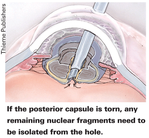
There are some more complicated instances in which the posterior capsule tears but there are still nuclear fragments present. "These situations usually occur when there's surge, and the capsule is caught up in the surge and is emulsified and torn by the phaco tip," remarks Dr. Fishkind. "The other setting is with a really hard nucleus, where sometimes the surge will cause the capsule to slap up against a sharp fragment of nuclear material and be torn by it. Both of these situations result in torn posterior capsules with or without vitreous loss and fragments still in the eye.
"The effort in cases of fragments is to isolate the fragments to keep them from falling through the vitreous hole," Dr. Fishkind continues. "When fragments and a posterior tear are present, I use a dispersive OVD, putting it over the rent in the capsule. I try to tell whether the vitreous is intact by watching how the OVD interacts with the hole. If the OVD seems to cover it smoothly and push things back nicely, then I can make the supposition that the vitreous is probably intact. But if the OVD goes in funny directions, doesn't cover smoothly or is hard to get to go where I want it to go, that's because there's vitreous holding it away. I then know the vitreous face is ruptured and there's vitreous in the anterior chamber to deal with."
Once the chips are isolated away from the hole, Dr. Fishkind puts OVD below the cornea in order to protect it. He then brings the fragments up to the plane of the pupil for phacoemulsification. "I'm trying to create as minimal a number of pieces as possible," he explains. "I want to keep them whole and not break them apart. If necessary, I'll add OVD to maintain the deep chamber and maintain the corneal protection." For a very large fragment, Dr. Fishkind won't hesitate to use a Sheets Glide to stabilize it during removal.
Once the nuclear fragments are removed, Dr. Fishkind advocates a manual removal of cortex, rather than automated removal with an I/A tip. "For me, manual removal involves a 25-ga. cannula on a 3-cc syringe that's about half-filled with BSS," he says. "Then, I grasp whatever it is I'm removing, such as cortex, aspirate it, take it out of the eye and irrigate it onto the drape. I just go back and forth doing that until I've got all the cortex out." A relatively old product that he says actually works well for this application is the Simco I/A cannula. "This is a dual-barrel cannula with one side hooked to irrigation and the other hooked to aspiration. So, you can irrigate through it and aspirate at your desired speed as the chamber fills with irrigating solution. The irrigation can be done either through the bottle or by an assistant who gently injects the fluid for you while you aspirate."
Zonular Instability
In cases of absent or loose zonules, surgeons say that being forewarned is forearmed. However, zonular problems can still occur without warning in some cases.
If there are zonular issues that you're aware of beforehand,
Zonular problems can also impact the nucleofractis technique you use. "If it's a firmer nucleus, I use vertical chop," says Dr. Whitman. "I can stay gently within the bag and keep the pieces from moving around within the bag. Also, I'm not using forces that push the bag this way and that, which would loosen the zonules further. Another option is pre-chop in which, prior to anything else, I elevate the nucleus into the anterior chamber.
Though I usually don't like working in the anterior chamber, this is a good situation in which to do it, because now I have the nucleus out of the bag. I'll pre-chop the nucleus with an Akahoshi pre-chopper or the like, phacoemulsify it, and then do a careful I/A and see what the bag's doing. Is it really pulling from somewhere? Do I see zonules pulling toward me as I aspirate cortical material? If not, I may be home free. If so, however, I stay away from those areas and clean everything else up. Then, I'll put in a capsular tension ring because I want to know that this is going to be stable not just for the injection of the IOL but also six years later. I'd like to know I've done everything I can to strengthen the zonular fibers and put less stress on them. The CTR pushes the bag out and puts it closer to the insertion of the zonules. Before I implant the lens I make sure the bag looks nice, round and symmetrical."
However, there are also cases in which there are three, four or five clock hours of zonules that are either loose or absent, or that were loosened during surgery. In this case, some surgeons say a CTR may not be the best option. You can sew a CTR segment to the scleral wall for stability and keep the lens in the bag, says Dr. Whitman, or you can just clean things up as best you can and either suture a posterior chamber lens to iris or sclera or implant an anterior chamber lens.
However, there are times when zonular problems happen unexpectedly. "A lot of times, we're doing our I/A and we go to remove some cortex and we see the whole bag pull toward us—not just the cortex—and we see a lot more striae in the bag. The bag will seem to pop up into the I/A very easily, behaving more like cellophane than a normal capsular bag," explains Dr. Whitman. "At this point, all the warning lights need to be flashing in your head. As soon as you see that, put in a CTR and you'll save the whole process. The CTR will usually stabilize the bag/zonular complex so you can put your lens in and tease out any remaining cortical material."
Iris Issues
Problems with floppy irises and small pupils can also complicate a case. Fortunately, surgeons have developed ways to counteract these problems and achieve good outcomes.
For patients on Flomax, some surgeons recommend preop atropine. "We prescribe atropine to be started three days prior to surgery and taken b.i.d.," says

Davenport
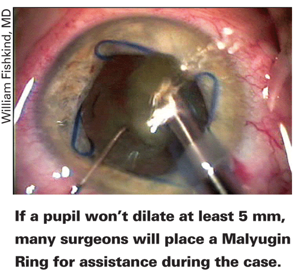
"This stiffens up most pupils. When people hardly dilate—either poorly or irregularly—in the office ahead of time and have a history of Flomax, then I'll use a combination of scopolamine, neosynephrine and mydriacyl for dilation during the case. I also put in the 1:4,000 epinephrine. If I don't have a 5-mm pupil after that, I place a Malyugin ring. If I do have a 5-mm pupil, I'll probably lay the peripheral iris down with Healon5 and pack that in with some Viscoat in a softshell technique to keep everything away from the cornea and the phaco tip. It's important to have perfect incision size and location, as well as taking care to maintain and not overfill the chamber."
"Inserting a Malyugin ring is a bit of a challenge, but getting it out is easy," adds Dr. Arbisser. "When putting it in, you first want to deepen the chamber with viscoelastic.
Then, you should be able to use a Sinskey hook through the paracentesis to help guide the distal loop into the right place to hold nasal iris as the ring emerges from the inserter inside the eye. Then, as you slowly let the other two parts emerge, you can usually capture those three loops with the superior, inferior and nasal iris. It's usually too much traction to push it enough to be able to then get the proximal loop in place without retracting the proximal iris, so I'll usually let the ring stay with just three of them in the right place. I'll just position the proximal one and put two instruments through the incision: a Lester hook to pull the iris back and a Kuglen hook to push the ring forward in order to capture the iris proximally. Also, make sure the incision is irrigated and sealed before removal of the ring."
Though surgeons say the ring comes out fairly easily, there are steps you can take to make it even easier.
"As you're getting the ring out, you have to disengage one of the loops," says Dr. Ferguson. "So, if you go in from the side with a Beckert nucleus rotator and lift that loop that's subincisional, sometimes it will go under the iris. All you have to do is rotate that ring and you've got another loop to work with, and now there's less tension on it. A lot of times people will try to fish for it, but the ring will rotate very nicely on the iris once one loop is disengaged."


