

Shortly after fertilization as the embryonic cells divide quickly going from two cells to 128 cells. This is called a blastula. The blastula has a spherical layer of cells called the blastoderm. The blastoderm consists of three layers—the endoderm, mesoderm and ectoderm—from the inside to the outside. The cells of each layer have distinctive characteristics that will enable the cells to develop into specific types of tissue. While we will examine only the eye, keep in mind that the same three embryonic layers give rise to all of the varied tissues in the body.
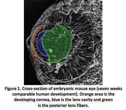
The human eye begins to develop during the 17th day of gestation. Mesoderm cells, the middle layer of the blastoderm, and ectoderm cells, the outer layer of the blastoderm, form the eye fields in the neural area of the embryo. Optic vesicles develop in the eye fields and in five days, infold to form the optic cup. At this point, the retina and crystalline lens begin to develop. Meanwhile, surface ectoderm cells are becoming thicker. The lens forms from these thickened cells. By the 32nd day, you can easily identify the lens, and during the next three to three and a half weeks the lens will grow to the size it will be at birth (Fig. 1).
 |
Between 30 and 35 days, you can see the start of the iris. In two more weeks, it is fully developed. While the iris grows, the optic stalk, the precursor to the optic nerve, forms a critical connection to the forebrain at 36 days.
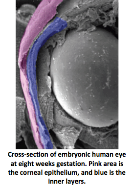
Eye development during the first trimester of pregnancy is like watching the grand finale of Fourth of July fireworks. Cells and tissue develop quickly and simultaneously to form various eye structures. For example, as the lens is developing, the cornea forms from ectoderm cells covering the lens. At about five weeks of pregnancy this tissue is two cells thick. What occurs over the next two weeks is amazing. The number of ectoderm cells will nearly double, creating the corneal epithelium (Fig. 2).
|
At this point, the lens has completed its formation and separates from the ectoderm. This separation creates the anterior chamber. Meanwhile, neural crest cells have separated from the neural plate and migrate to the anterior chamber to form other neuronal and non-neuronal cells. Once separated, they move into the area between the lens and the corneal epithelium, forming the corneal endothelium and stromal keratocytes, the primary cells of the stroma. To all that activity, add the formation of the orbits and extraocular muscles at four weeks gestation. Depending on eye growth after birth, however, the orbits may not mature until adolescence.
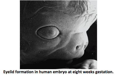
During the sixth week the lacrimal glands begin forming but produce no tears until the third month after birth, which is why infants shed no tears when they cry. At eight weeks, the eyelids start to form and fuse together protecting the other developing eye structures (Fig. 3).
|
During the fifth month, a two-month process begins—the separation of the eyelids.
While the eyelids are fused, the corneal epithelium decreases back to two cells in thickness and enlarges and matures. The maturing, highly hydrated cornea does not become transparent until the eye becomes functional in the seventh month. Descemet’s membrane matures just prior to the eyelids opening. Bowman’s membrane develops in the fifth month, as the cornea becomes innervated.
Although the iris begins to develop from the rim of the optic cup at five weeks, the muscles controlling the aperture do not develop until five months. Since constriction of the pupil in response to light is not necessary in the darkness of the womb, it doesn’t begin until the eighth month. It takes another two weeks for this response to become consistent. Because this reflex develops late in gestation, babies born prior to 34 weeks gestation need eye protection.
In a neonatal intensive care unit, for example, premature babies wear eye covering. In the 1970s and 1980s, the medical community believed that stimulating visual attention, focus and tracking was critical to the visual development of premature infants. Recent research, however, shows that because the light control response is underdeveloped, the need for protection from light outweighs the need for visual stimulation until the premature infant has reached the point in development that would have been complete at 40 weeks gestation.
At seven weeks, the sclera develops from embryonic tissue, which allows the formation of blood vessels. The cornea connects to the sclera, which is nourished by blood. But why then does the cornea lack blood vessels? The answer is that the cornea develops from ectoderm rather than embryonic tissue. The ectoderm cells provide a clear medium for light, and the lack of blood minimizes tissue rejection in corneal transplants. It is for this very reason that the cornea forms from the same embryonic cells as the crystalline lens. How’s that for efficiency? In addition, we also know that both tissues have refractive power.
During this time the retina is still evolving. The neural cells of the retina develop from a single embryonic layer. The cells then differentiate into specific types according to their projected role in the visual process, such as rods and cones, with axons from the retinal ganglion cells forming the optic nerve. It takes six months for the retinal layers to grow from the neural ectoderm. The macula needs four or five months just to begin, and it will mature six months after birth. As the retina and lens develop, the vitreous forms between them. In the meantime, the neural connections between the eye and brain have been developing, taking five months to complete.
The bulk of the highly complex development of the retina occurs between 24 weeks gestation and 3 to 4 months of age, when the optic nerve becomes fully myelinated. Rod cells are better developed at birth resulting in scotopic vision in infants. As a result, newborns see primarily dark, light and shades of gray. Color discrimination occurs at 3 months of age when the cones mature. Between 28 and 30 weeks gestation, the fetus develops rapid eye movement and sleep patterns. What does that have to do with vision?
The connection between the retina and the brain is the essence of vision, and REM and sleep cycles help messages from the retina synchronize with the brain waves of the visual cortex. If these cycles are disrupted or the infant is deprived of sleep, it can significantly interfere with visual development.

