


This year's ARVO abstracts shed light on a number of key issues, including the connection between dry eye and contact lenses, techniques for evaluating the impact of contact lenses on the cornea, new ways to visualize the tear film, new evidence regarding soft contact lens wear and myopia, and risk factors for severe microbial keratitis in daily wear contact lens users. (Unless specified, studies had no commercial support.)
The Dry-Eye Connection
Meibomian gland dysfunction has been recognized as a possible cause of dry eye in contact lens wearers. With that in mind, researchers in
Using their new technology, they examined one eye of 121 contact lens wearers (39 wearing gas permeable lenses, 82 wearing hydrogel lenses) and one eye of 137 age-matched non-contact lens wearers as a control. Among the contact lens wearers, the period of wear was 12.3 ±7.2 years and the spherical equivalent was -5.1 ±2.7 D. The status of their meibomian glands was scored as follows: 0=no loss of meibomian glands; 1=less than one-third of total area lost; 2=between one-third and two-thirds lost; 3=more than two-thirds lost. (Scores in upper and lower eyelids were summed for each subject.) Subjects also received a questionnaire, manifest refraction, slit-lamp exam, conjunctival staining with fluorescein, measurement of tearfilm breakup time and Schirmer I test.
The results indicated that contact lens wearers have significantly higher meibomian gland loss than age-matched non-contact-lens-wearers:
• Scores were significantly higher in wearers (mean: 1.71) than among controls (mean: 0.96) (p<0.0001).
• Average contact lens wearer score was similar to that of 60- to 69-year-olds in the normal population.
• Length of time wearing contact lenses and meibo-score were significantly correlated (p=0.0032).
In addition:
• TFBUT was significantly shorter in contact lens wearers (4.8 ±2.6 seconds) than in controls (6.7 ±3.7 seconds) (p<0.0001).
• Schirmer values in wearers and controls were 20.4 ±10.1 mm and 20.2 ±11.2 mm, respectively (p=0.96).87
Practitioners at Hazleton Eye Specialists in
After a two-week washout with Refresh Tears q.i.d., all patients were fitted with AA lenses OD and either PB or EE lenses OS. Half of each group used Refresh Tears as needed, while the other half used Restasis in both eyes b.i.d.—no artificial tears allowed. During follow-up visits at week one and months one, three and six, the two sites measured Schirmer scores, TFBUT and patient assessment of lenswear time and comfort.
The data showed that all the new lenses outperformed the previously worn lenses for all outcome measures by the one-month exam (p≤0.001). PB and EE lenses performed about equally well, but both outperformed the silicone hydrogel lens (AA) for all outcome measures by three months (p≤0.007). In all cases, Restasis produced significantly better outcomes than artificial tear use on all measures by one month (p≤0.009).101
Researchers at the Institute for Eye Research in
Using data gathered at the two-week, one-month and three-month visits, plus a two-month questionnaire, they compared participants who discontinued the lenses to those who did not. (They disregarded data from any subjects who discontinued because of an adverse event.) In the group that discontinued use:
• A greater proportion were less than 20 years old (p=0.027) and were new to SiHy lenses, or contact lenses in general (p=0.001).
• They reported a higher frequency of moderate-to-severe blurred vision, discomfort, lens awareness, burning and stinging, dryness and redness (p<0.03).
• They fared worse in terms of self-reported redness, itching and overall awareness of the lenses (p<0.01).
• They rated their lenses lower for overall comfort, comfort on insertion, comfort during the day, comfort at the end of the day, ease of lens handling, overall vision, overall dryness and end-of-day dryness (p<0.01).
• They reported shorter hours of wear (p=0.001) and comfort (p=0.000).
After eliminating adverse-event-related subjects, the discontinuation rate was 10.7 percent in the first three months. (Only 4.5 percent discontinued because of an adverse event.)4839
Visualizing the Tear Film
Two doctors at the Bascom Palmer Eye Institute in Miami conducted a pair of studies using a slit-lamp-based ultra-high resolution optical coherence tomographer (SD-OCT) developed with advanced optical delivery, 3-µm depth resolution and scan width up to 15 mm, to visualize the tear layer and contact lenses on the ocular surface in vivo. (One doctor has received funding from Allergan and Bausch & Lomb.)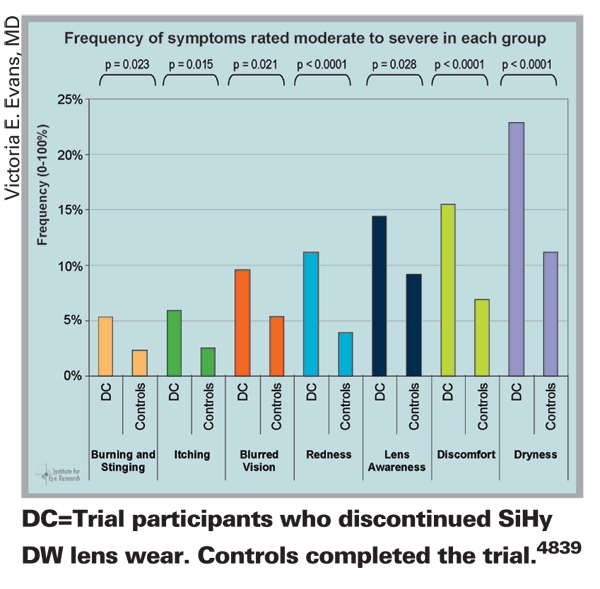
The first study demonstrated the feasibility of this approach. The study authors imaged the eye before and after wearing both soft and hard contact lenses, as well as following instillation of a drop of artificial tears. They were able to directly visualize the tear film in both normal and dry-eye patients, including the tear menisci around upper and lower eyelids, lens edge configurations, the tear meniscus around the lens edge, and the pre- and post-lens tear films at the lens edge. They also imaged the course of tear thinning over time after blinking, and obtained images of corneal layers such as Bowman's and the epithelium.5310
In the second study, the authors used the SD-OCT scanner to acquire high-quality 3-D images of the cornea while using Intralipid, a biocompatible optical scattering medium, to enhance the boundaries of the tear meniscus and contact lenses. When scanning a contact lens on the eye following instillation of artificial tears alone, the boundary between the artificial tear and the contact lens was clear only at the central portion of the lens; the boundary between the tear meniscus and the contact lens was not recognizable. With Intralipid, boundaries between the contact lens and the upper and lower tear menisci could be clearly visualized, and both surfaces of the lens could be seen clearly across the entire image range. (See images, below.)
The authors note that with the boundaries of the tear meniscus enhanced, it should be possible to calculate the volume of the tear meniscus—an important parameter in the diagnosis and study of dry eye.4267
Contact Lenses and the Cornea
Researchers at
The CL-wear groups both had significantly lower mean CH and CRF than the controls. (Differences between the one-day and two-week groups were not significant.) No significant differences in CCT were found among the three groups, although there were significant correlations between the CCT and CH (p< 0.05) among three groups.)661
Researchers at the Southern California College of Optometry and Alcon Research conducted a randomized, concurrently controlled, double-masked study to determine corneal responses to different contact lens material and care-solution combinations. To determine bio-incompatibilities, they used the single-drop epithelial barrier function method,1 as well as corneal staining.
Nineteen young, adapted, flexible daily lens wearers were used as subjects. New silicone hydrogel or hydrogel lenses (galyfilcon A and omafilcon A, respectively) were soaked in pre-conditioned lens cases in either a solution containing polyquaternium-1, a PHMB-based solution or non-preserved saline (the contralateral control). Lenses in the multipurpose solutions were soaked six to eight hours (test condition); control lenses were soaked for two hours (active control).
After two hours of wear, researchers used the single drop method and a scanning fluorometer to determine epithelial barrier function and calculate the fluorescein penetration rate (Pdc) in nm/sec. Corneal staining was also assessed immediately following barrier measurement.
The mean Pdc ratios (test solution to control solution) were: PHMB/galyfilcon: 2.1; PHMB/omafilcon: 9.6; polyquaternium-1/galyfilcon: 1.9; and polyquaternium-1/omafilcon: 1.2.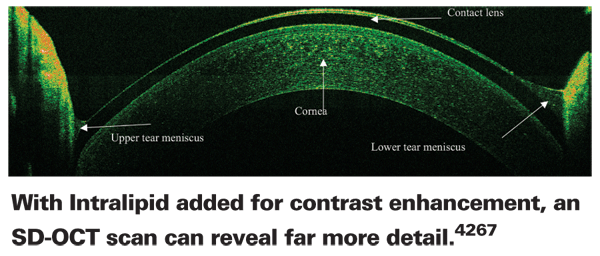
The only statistically significant difference was between the PHMB/ omafilcon-A combination and polyquaternium-1/omafilcon-A combination (p=0.001). In addition, corneal staining scores paralleled the Pdc ratios. For example, the PHMB/omafilcon-A combination produced a Pdc ratio of 9.6, reflecting a more than 900-percent increase in penetration rate of test vs. control; median corneal staining in the test eye was grade 3, while the control was grade 1.5.
The researchers conclude that large differences in lens-solution bio-incompatibilities relative to corneal compromise can exist, which may be mirrored by corneal staining data. They also note that the single-drop barrier method appears to be a useful means to obtain sensitive, objective evidence of corneal compromise.2019
Researchers at the
Both CCT (+35 ±8.8 µm) and IOP (+2.4 ±1.2 mmHg) increased significantly after lens wear (p<0.001). The study authors conclude that a relatively small increase in CCT from contact-lens-induced corneal edema caused an overestimation error in Goldmann tonometry measurements. However, there was no correlation between the increases in IOP and CCT; and although IOP returned to pre-lens-wear values 40 minutes after lens removal, CCT remained significantly thicker than baseline 60 minutes after lens removal (p<0.001).690
Causing Myopia Progression?
Because many previous studies of contact lens wear and myopia only lasted a year or less, researchers at five different colleges of optometry collaborated in a study that followed 484 8- to 11-year-old children for three years. (All authors receive funding from Vistakon.) In this study, 237 of the subjects were randomly assigned to wear glasses; the other 247 wore soft contact lenses (Acuvue 2 or 1 Day Acuvue). Researchers measured refractive error, corneal curvature and axial length, prior to randomization, and then annually.
• Spectacle wearer progression was -1.08 ±0.71 D, while the contact lens wearers progressed -1.27 ±0.72 D (covariance, p=0.005).
• The axial growth of spectacle wearers was 0.59 ±0.37 mm; axial growth for contact lens wearers was 0.63 ±0.34 mm (p=0.27).
• Change in steep corneal meridian was 0.05 ±0.69 D for spectacle wearers and 0.10 ±0.70 D for contact lens wearers (p=0.43).
Although three-year myopic progression was statistically greater for contact lens wearers, the study authors note that the difference was only 0.19 D, which is not clinically meaningful.2021
Contact Lenses and Infection
Researchers at multiple sites in
Cases and controls were interviewed by phone to determine details of lens type, wearing habits and history, and demographics. Researchers used a clinical case definition and stratified cases by severity according to the size and position of the lesion, culture result and visual outcome. During the study period, 125 eligible cases were reported, including 90 cases of severe keratitis; researchers identified 1,090 community controls wearing daily wear contact lenses.
The data showed that, after adjusting for age, gender and lens material type, independent risk factors for severe keratitis infection included: occasional overnight lens use (once a week or more often—6.5x); high socioeconomic status (4.1x); poor case hygiene (6.4x); smoking (3.7x); infrequent lens case replacement (5.4x); and solution type (7.2x). Neither lens material, wearer age nor gender was associated with severe disease.4853
Researchers in Alcon's Microbiology research and development division tested a new method for evaluating the effectiveness of a contact lens disinfecting solution against Acanthamoeba. (Currently there are no standardized methods for accomplishing this.) Their methodology was based on the U.S. Food and Drug Administration's, American National Standards Institute's and International Organization for Standardization's stand-alone and regimen tests for bacteria and fungi, modified to work with Acanthamoeba. Opti-Free RepleniSH Multi-Purpose disinfecting solution, a no-rub solution that contains polyquad and aldox preservatives, was challenged with cysts and trophozoites (trophs) of A. castellanii ATCC 30234 and A. polyphaga ATCC 30871. The stand-alone test used cysts (the more resistant form of the organism); the regimen test used cysts and trophs.
Three lots of RepleniSH were evaluated in the presence of organic soil:
• In the stand-alone test, a volume of RepleniSH was inoculated with the challenge organism. At disinfection time, an aliquot was removed, diluted and plated using a quantitative mirotitre plate method.
• In the regimen test, silicone hydrogel lenses were inoculated with the challenge organism; the no-rub regimen was performed and each lens was placed in a lens case containing about 4 mL of test solution. At disinfection time each lens and a sample of the lens case solution were diluted in microtitre plates.
Viable organisms were recovered with E. coli ATCC 8739. Plates were observed for viable amoeba after 14 days using an inverted microscope; survivors were enumerated. Stand-alone test results showed a 2 ±0.3 log reduction for A. castellanii and a 1.6 ±0.7 log reduction for A. polyphaga cysts at disinfection time; regimen test results showed no survivors for 95 percent of the lenses. Fewer than 10 survivors per lens were observed on the remaining lenses. (The study authors note that these methods are a reliable and objective way to evaluate lens solutions against Acanthamoeba.4871
Lens Wearer Preferences in
To help generate a picture of current contact lens-user preferences in
The most popular lenses were two-week frequent-replacement lenses, purchased by 49.9 percent; second most popular were daily disposable lenses, purchased by 36.4 percent. (Even though lens use decreased in those age 40 and over, the number of daily disposable lens wearers remained relatively high. Mean user age for these lenses was 30.9 ±10.2 years.)
Ninety-four percent of lenses purchased were soft contact lenses. Two-week lenses tended to be more popular with younger wearers. Lens type purchased were: spherical, 83.8 percent; toric, 10.8 percent; color, 4.7 percent; bifocal, 0.6 percent. Also, the majority of the subjects (52.6 percent) used multipurpose solutions in their lens-care routines. Hydrogen peroxide was the second most popular choice (27.3 percent).4841
1. Fahim MM, Haji S, Koonapareddy CV, Fan VC, Asbell PA. Fluorophotometry as a diagnostic tool for the evaluation of dry eye disease. BMC Ophthalmology 2006;6:20.


