


While many surgeons choose to perform PRK after RK, I first prefer to use LASIK as a secondary refractive procedure if the patient is a good candidate. This reduces the incidence of postoperative haze, while preserving the use of PRK for subsequent surgeries that may be required in the future due to progressive hyperopic shift and cataract surgery. In this article, I'll explain my reasoning and my approach to LASIK in post-RK patients.
Some History
In 1990, I first began performing RK and PRK. I was in the Food and Drug Administration-monitored PRK trials and the LASIK trials. I was also part of the first study of PRK over RK conducted in
Then mitomycin came on the scene. It was designed and introduced to be used in PRK to lessen the incidence of haze. I use mitomycin in select cases and feel it has been an important addition to cases where surface ablation is the best choice and where haze is a significant issue. However, the long-term effects of mitomycin are still unknown. As such, patients deserve to know their options with regard to secondary refractive procedures, such as PRK and LASIK. When given the choice, some patients might choose to have the risk of a flap to lessen the risk of haze or a potential long-term mitomycin complication. Others, after learning about the risk of the LASIK flap, might choose to take the mitomycin route and go with the PRK procedure. As surgeons we should discuss the options fully with patients so they can make a well-informed decision about their options.
Consequences
It is helpful to divide these decisions into short-term and potential long-term consequences. As mentioned, with PRK over previous corneal surgery, like RK, there is an increase in the incidence of haze. Further, the incidence of haze rises with every PRK procedure that is done after RK. For example, a 46-year-old patient may do well with his PRK over RK. In 10 or 20 years, the patient may need repeat surgery because he has had a hyperopic shift from the RK. Now the surgeon must discuss PRK over PRK over RK with a second dose of mitomycin (or another newer antimetabolite drug that may be available). The risk of haze goes up with each corneal surgery.
As a surgeon, I always consider the long-term situation, and there are several factors to take into consideration. I prefer to keep the PRK for the smallest correction possible because it is known that with less tissue removal, there is less risk of haze. I also like to keep PRK in the patient's armamentarium as a long-term enhancement option.
Therefore, if possible, I start with LASIK. The LASIK procedure brings the patient's refractive error closer to
Long-term Situations
Progressive hyperopic shift and cataracts are two of the long-term situations the surgeon will want to consider. If the patient has a progressive hyperopic shift, the surgeon will still have PRK as an option to enhance him if LASIK was performed previously.
Also, with time and advancing age, these patients could develop cataracts. While I do not see patients with previous refractive surgery experiencing cataracts at a higher rate, I do see early cataracts bothering this patient group more than those patients who have not had refractive surgery. A multifocal cornea from previous refractive surgery—especially RK—in combination with an early cataract can degrade image quality much more quickly than the same early cataract in someone who hasn't had previous refractive surgery. Therefore, surgeons have to keep in mind that cataract surgery is in the future of many of these patients. In addition, because the implant calculations after previous corneal surgery are not as accurate, it is a good scenario to have the option of another corneal refractive surgery after cataract surgery.
Another concern is that when PRK is performed, the epithelium thickens, and there is epithelial hyperplasia. For instance, with the flap maker, surgeons have to take into account that the epithelium is somewhat thicker, and making a flap after PRK can be quite challenging. There are risks involved, and that is why I like to prevent that. Therefore, if my patients were going to need all three procedures in the long run—PRK, LASIK, and cataract surgery or even just PRK and LASIK—as the surgeon I would definitely want the order of events to be: first, LASIK; then the PRK or cataract; and third, the PRK (if the patient has had previous cataract).
Additional Considerations
There are also several special nuances to consider. If the patient has large or significant epithelial plugs in the incision, he is most likely not a good LASIK candidate because the epithelial plugs greatly increase the chance of epithelium growing under the flap. As we know, one of the dreaded complications of LASIK after RK is the epithelium growing under the flap; this can be difficult to stop. In these situations, the informed consent becomes very important with the patient. If the patient has recurrent epithelial ingrowth under the flap, there is the risk that cleaning out that epithelium and suturing that flap is not going to prevent that recurrence (even though it should). In some cases, if epithelial ingrowth under the flap continues to recur, flap amputation or removal may have to be performed. 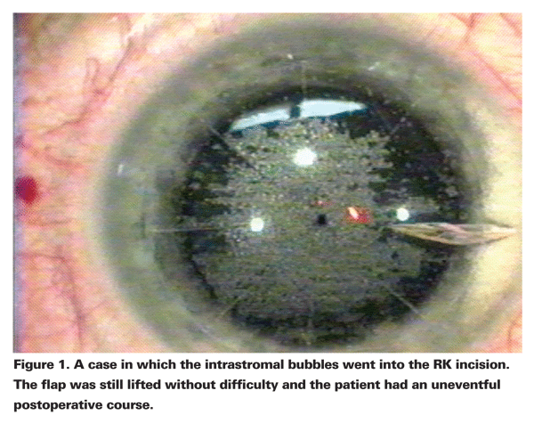
That said, I have done LASIK after RK 120 times, and I have not ever had to amputate a flap. I have had to suture the flap in two cases to get rid of recurrent epithelial ingrowth. While that is something that can happen, I have also dealt with corneal haze following PRK after RK that has bothered the patient and I much prefer that to dealing with epithelium under the flap. This is because the haze after previous corneal surgery can be very debilitating to the patient.
Therefore, I do not like to do LASIK in flaps with significant epithelial plugs. It is important for the incisions to be thin, well-healed and at least two years postop. In addition, previous ocular surface disease, such as dry eye, should already have been aggressively treated. I have a low threshold for turning to punctal plugs, Restasis and bedtime ointments. I also counsel patients to make sure they don't use a ceiling fan or anything that can dry their surface so that their healing is maximized.
On occasion, I will lift up a flap and enhance. I take this decision very seriously. I tell my patients that even though I traditionally like to wait six months for enhancement decisions, occasionally I will consider an early enhancement at three months if there is significant refractive error, and the flap performed beautifully with the first LASIK procedure. This is because early on, at just three months, I can oftentimes lift that flap without separating the incisions because that is the goal.
Flap Creation Technology
For LASIK as a secondary refractive procedure after RK, there is also the issue of using a bladed microkeratome versus a femtosecond laser to make the LASIK flap. I always advise patients that if my incisions were healed and sealed, I would chose to have the femtosecond flap. However, I also explain that they need to realize that if, during the lamellar dissection, the intrastromal bubbles escape through the RK incision and get between the corneal epithelium and the focusing lens of the laser, then the procedure will need to be stopped. This is because when the gases and fluids get between the epithelium and the focusing lens, they block the laser and the flap is not made (or an irregular flap is made).
If that situation occurs, I would have the patient return in six months and give him the option of a microkeratome blade flap for LASIK or PRK with mitomycin. In my 120 cases, this has happened twice. The gases did come through the incisions and I stopped the procedure. In both situations the patients returned six months later and received blade LASIK flaps, and they did very well.
If a patient has epithelial plugs but they are not very impressive, meaning there is not impressive incision gaping, and he still would like to have LASIK, I would lean toward the microkeratome flap rather than the laser flap, knowing that in those areas of epithelial plugs that the breakthrough can happen with the bubbles.
If a patient has significant epithelial plugging, oftentimes the incisions are gaping. These patients have a tendency to be worse candidates for PRK, too, because they tend to haze more aggressively. These issues must be part of the informed consent. I do not make flaps in those situations. This can be a tough decision. If the patient's correction is not too high, I would lean toward PRK. However, first I would make sure the patient had failed gas permeable contacts and he is not satisfied with his spectacle correction. I would also ensure that the patient understands the situation and its potential outcomes.
As far as intraoperative suggestions, during the flap creation, I prefer to make a thicker flap than in a routine case. The goal is to minimize the chance of the intrastromal bubbles tracking through the incision and getting between the focusing lens and the cornea. Thus, I like to use a 130-µm minimum laser flap. I also like to use this thicker flap minimum for blade flaps because, whether the flap was created with a laser or a blade, the more robust flap can help minimize the chance of incisional separation. In Figure 1 you can see an Intralase-created (AMO) flap where the intrastomal bubbles tracked into the incision but did not break through; thus the flap was still lifted and the laser correction performed without difficulty.
Flap-Lifting Technique
I have found that this flap-lifting technique done with extra care and patience can be very successful. With the IntraLase flap, the flap may lift very smoothly or there may be microadhesions that the surgeon needs to separate before the flap can be lifted. Typically when I am doing flap lifts with the Intralase, I do not fixate the globe. However, I do fixate the globe with .12 forceps when I am lifting laser flaps over RK because I am attempting not to stress or separate the incisions. Any micro-movement from the patient could also affect this process.
First, with a Slade Refractive Spatula (Bausch & Lomb/Storz), I separate the micro-adhesions at the edge of the flap between the incisions (See Figure 2). I take care to not swipe across the junction of the incision and the flap edge. Instead, in the flap periphery, I separate the areas where there is no incision first.
Then I go under the flap, in between the incisions, with the Slade spatula and go to the center. Then, at the inner aspect of the incision, about 1 mm, I orient the Slade spatula so it is perpendicular to the incision as I swipe under the incision. I have my Slade spatula with its peripheral aspect a couple of millimeters past the incision to ensure that I do not put any undue stress on either side of the incision. As I swipe I make sure that my Slade spatula is fully underneath it and then I take it out to the periphery and separate the incision. In the majority of cases, if I put equal stress on both sides of the incision, they do not separate.
Next, I proceed around and I separate each incision in that way, one at a time. When I have all the incisions separated, I do one final swipe of the whole interface to make sure all adhesions are separated. Then I lift the flap with the Slade spatula and perform the ablation. I then lay the flap down and rinse, and smooth the flap into position as I routinely do.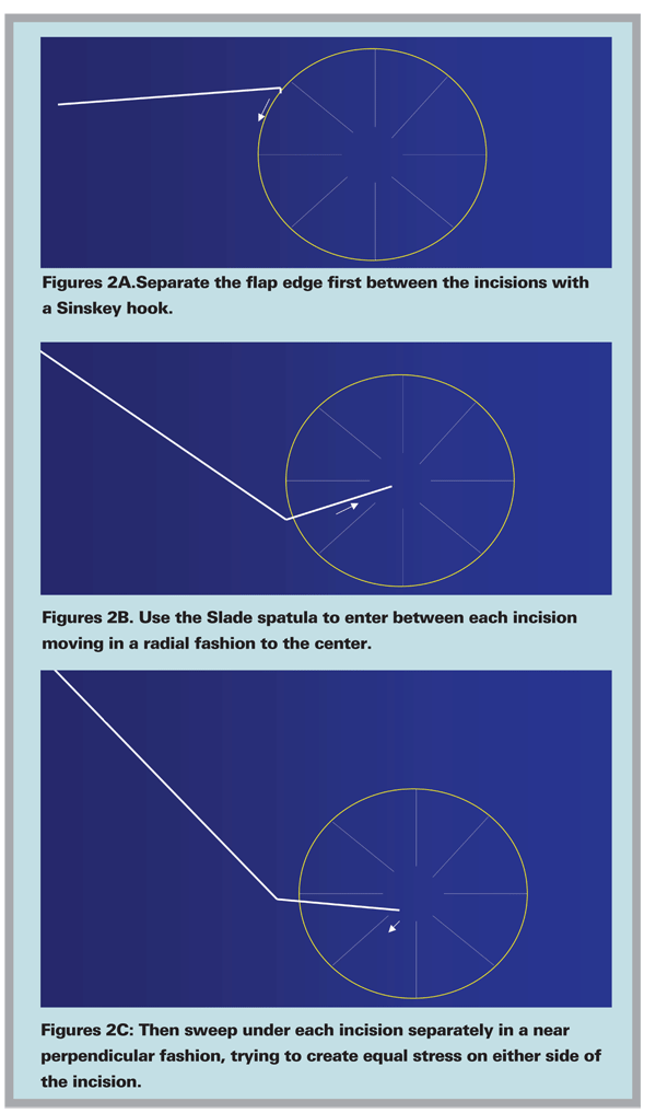
It is important to use corneal marks on an eight-incision RK marker prior to lifting the flap, because these incisions should line up perfectly. Incisions are not the easiest to see under the operating microscope, therefore the marks placed before lifting the flap become crucial.
In general it is best to never lift those flaps again. Occasionally, an enhancement is needed if there is significant refractive residual error. One option is to lift the flap and laser. I would use the same technique going in on the edge and swiping underneath each incision. However, I do not want to wait until that interface is so well-healed and sealed that I would put a great deal of stress on these incisions. Thus, three months postop is a good time for this discussion. The patient needs to be fully aware of the increased risk of epithelial ingrowth if this rare decision to lift the flap is considered. In addition, I advise these patients that if, in that first entry under the flap there is much friction, I would cancel the procedure and explain that we are not even going to attempt it.
In general, I consider LASIK after RK a one procedure event—one flap—and everything has to come together perfectly for me to consider lifting it a second time. By this I mean there is enough residual refractive error that I would prefer not to use PRK and the incisions are thin and well-healed with no epithelial ingrowth. Only then would I go in to relift the flap, and only if there is not much residual friction to complete the procedure.
Overall, I have had very good experiences with laser flaps after RK. I have been very satisfied with the procedure, and I think it is worth considering so that patients can preserve their refractive options over the course of their lifetime.
Dr. Thompson is the director of Refractive Surgery for the Sanford Clinic in


