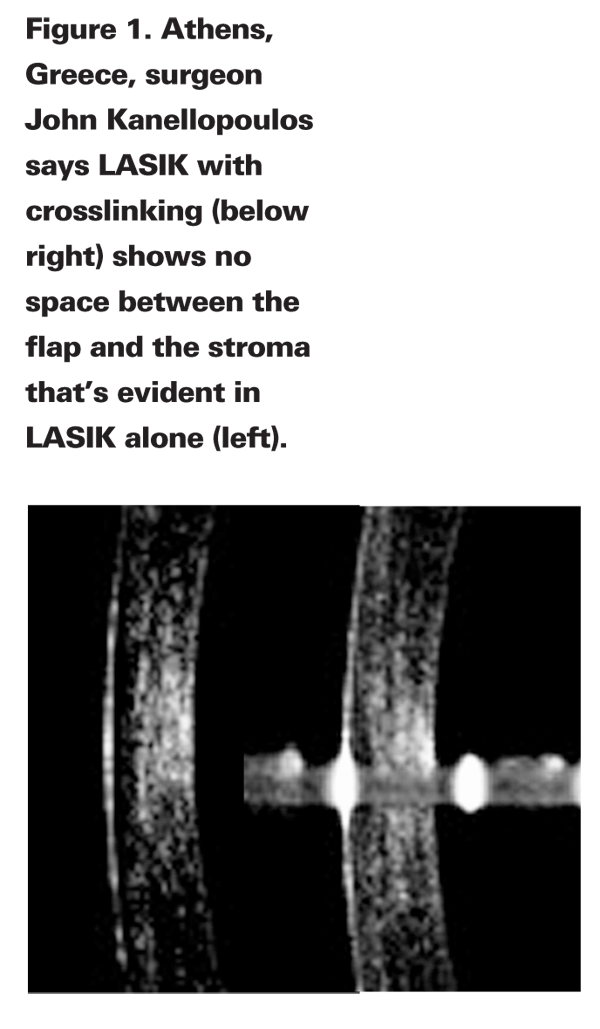
With the current surgical atmosphere emphasizing avoidance of ectasia at all costs, some refractive surgeons may be surprised to hear that LASIK is being performed specifically on corneas that aren't very thick or that have unusual topographies.
Treating Risky Eyes
Dr. Kanellopoulos says performing LASIK on eyes he describes as "high-risk" wasn't a decision that was made overnight, but instead came as the result of an evolution of his use of corneal crosslinking with riboflavin and UV light.
"Since 2002, we've treated several cases of post-LASIK and post-PRK ectasia with the standard crosslinking treatment in which we remove the epithelium and instill the riboflavin drops on the surface, and we published several papers on it," says Dr. Kanellopoulos. "At the AAO meeting in 2007 we presented on a new modality of crosslinking in which we use the femtosecond laser. In this method, we place riboflavin directly into a specific level of corneal stroma in a novel administration process. Then, as a third step in the thought process, throughout the last six or seven years we've encountered some early LASIK ectasia cases in which we've employed this technique because of the significant reluctance of the patients to undergo epithelial removal by way of the classic crosslinking method. We encountered two or three cases in which there was enough corneal tissue reserve to attempt an intrastromal crosslinking without removing the epithelium. After seeing it work in some of these mild post-LASIK ectasias that had enough tissue in reserve to do an enhancement procedure and additional crosslinking, we've employed it prophylactically over the last two years in a select group of patients, the high-risk LASIK cases, with very gratifying results."
When Dr. Kanellopoulos refers to a high-risk LASIK patient, he means eyes in which the initial corneal thickness isn't optimal. "This would be where someone is starting with a corneal thickness of 500 µm," he says. He says it might also be a patient who, regardless of his initial corneal thickness, is going to undergo a significant amount of myopic correction accompanied by a significant amount of tissue removal, such as a -8 D myope with a 510 µm preop corneal thickness. "I think this would be anyone who has funny-looking topography in which there's any question of posterior corneal elevation or an irregularity on surface topography and we conclude it's not forme fruste keratoconus," explains Dr. Kanellopoulos. He draws the line, however, at performing LASIK on corneas under 480 µm in minimal thickness.
The prophylactic treatment is about half of a standard crosslinking treatment, says Dr. Kanellopoulos. After the flap has been made with the IntraLase laser and the cornea ablated, he puts several drops of riboflavin directly onto the ablated stromal bed and the backside of the flap. Flap thicknesses vary between 100 and 120 µm. He waits a few seconds for the agent to be absorbed, and then lays the flap back onto the bed without pushing the fluid out. He waits another 15 to 20 seconds. "Within seconds, the flap is colored yellow, indicating the riboflavin has penetrated the totality of the flap and has soaked some of the underlying stroma," he says. At that point, he irradiates the cornea for 15 minutes with the standard crosslinking energy dose of 370 nm UV light with a fluence of 3 mW/cm2.
"With this technique, we're primarily targeting the actual link between the flap and the underlying stroma," Dr. Kanellopoulos says. "There was evidence presented by 
In a study of his technique that he presented at last year's AAO meeting, he presented data on 25 cases. The average follow-up was 1.5 years (range: one to three years). The spherical equivalent of the patients was reduced from an average of -7.5 to -0.2 D, and the average K reading went from 44.5 D to 38 D. The average flap thickness was 105 µm, and the average corneal thickness was reduced from 525 µm to 405 µm. The mean endothelial cell count went from 2,750 to 2,800. Dr. Kanellopoulos says the corneas are currently stable.
The Best Option?
Since Dr. Kanellopoulos' study involves high-risk LASIK eyes, surgeons familiar with crosslinking have varying opinions on the treatment.
"[Questioning LASIK in such eyes] is a valid criticism," says the Cleveland Clinic's director of refractive surgery and crosslinking researcher, Ronald Krueger. "A surgeon might say, 'Wait a minute, you're taking a higher-risk patient that we'd normally be canceling out of possibly treating and treating him?' Such an investigation might be viewed as experimentation and some might think it shouldn't be done. But, if you're in a place where you really want to do something to help a patient and, at least on a limited investigational basis want to do a series of patients that are fully informed, you might be able to justify that this can be done as long as you follow proper protocols. It might need institutional review board approval or something along those lines.
"I like the concept if it's really proven," Dr. Krueger continues. "If we can do it in a proper clinical trial and we demonstrate that it's safe and effective, I like the concept in the sense that we have patients who have every desire to have the best correction they can get but who are limited for some reason. It would be an exciting expansion of our field. However, in the
"Such questions are good ones," says Dr. Kanellopoulos. "If a patient's tissue reserve is sufficient, and the topography and tomography isn't in the red as far as signs for ectasia, and the patient's age is acceptable—with younger patients being more prone to ectasia—I think I'd feel pretty comfortable treating a patient. We haven't treated extreme patients, but I think it's made us feel much more comfortable in treating high-risk patients."
Some surgeons may question why LASIK is even necessary, when another procedure might do, instead. "Based on the study of the technique, we know these are high myopes to begin with, with an average preop sphere of -7.5 D," says
Dr. Kanellopoulos argues, however, that LASIK, especially if combined with crosslinking, is superior to PRK with crosslinking. "LASIK and crosslinking offers some significant advantages," he says. "Obviously, the postop recovery time and morbidity that the patient undergoes is worse with PRK. And there's less of a chance for scarring with LASIK, since coupling PRK with crosslinking can involve corneal scarring … There's always the argument that perhaps a partial PRK with crosslinking and a phakic IOL, or just a phakic IOL alone, might be a more stable, less risky approach. I think only time will tell what method is the safest, most effective one."


