
By Thomas H. Clark, O.D.
UNDERSTANDING ANGLE KAPPA
Part 2 discussed the effect of angle Kappa on PAL dissatisfaction. Part 3 defines angle Kappa and shows its relationship to fitting issues.
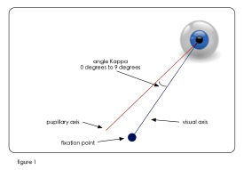
The accepted definition for angle Kappa is:
1. The angle between the pupillary axis and the visual axis. Termed positive when the pupillary axis is nasal to the visual axis, and negative when the pupillary axis is temporal to the visual axis (See Figure 1).
The biometric issue of angle Kappa is prevalent in almost all our patient population and is not an isolated issue as is shown in this study graph (Fig. 2).
The Journal of Refractive surgery published a paper of 800 tested eyes (Hashemi et al, Jan 2011). It showed the prevalence of angle Kappa differences with a range of 0 to 9 degrees with the mean angle kappa being 5.56 degrees. (2)
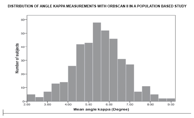
A 5.56 degree angle kappa error would result in a linear error of approximately 2.0 mm at the lens plane in a patient’s PAL RX.
The Fitting Issue
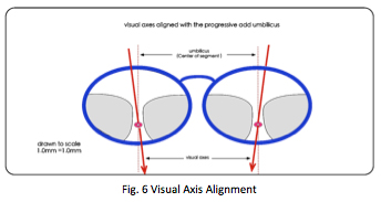
No one would argue that for a progressive add lens to be successfully fitted, the visual axes of a patient must intercept the PAL umbilic at the lens plane (see Figure 3).
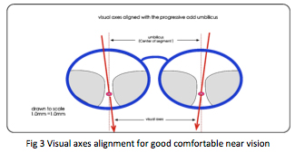
Aligning the PAL segment’s umbilic with a patient’s visual axes assures best vision. The parafoveal and foveal cone of vision will be centered along the umbilic within the PAL’s corridor and near (Fig 3). Misalignment would cause the cone of vision (parafoveal and foveal) to view through the distorted zones outside the progressive channel, resulting in blurred distorted vision.
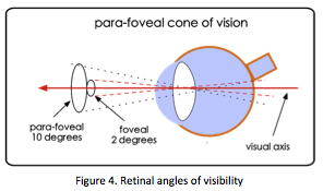
The Parafoveal Cone of Vision
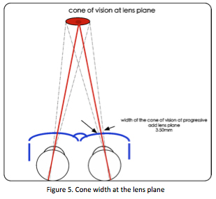
The foveal cone of vision is ~2 degrees while the parafoveal cone of vision is ~10 degrees (see Fig. 4).
At 10 degrees, the width of the parafoveal cone of vision, intercepts the umbilic of a progressive segment at a measured width of approximately 3.5mm (Fig 5). This 3.5mm circle at the lens plane falls completely within an intermediate zone of 5mm. This only works when the visual axis intercepts the umbilic of the patient’s progressive segment (see Figure 6).
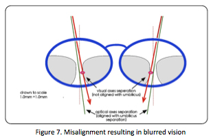
When the visual axes and the umbilic of the progressive segment are misaligned, the optical axes can be perfectly aligned but there will be blurred images in one or both eyes. This is caused by the resulting visual axes falling outside the clear zones of vision within the corridor and near. The vision cone crosses over transition areas of blur, present in all progressives, see Figure 7.
Part 4 will discuss the effect of the dominant eye on alignment error.
Dr Thomas Clark OD, http://www.willistonoptometrist.com/
For further information on this measuring system contact iPCapital Group