


| |
Volume 5, Number 17
|
Monday, May 2, 2005
|

|
||
Editorial: Learning
From Our Past
Fortunately, the past ASCRS meeting in Washington, D.C., was not one of those events. Certainly the World Cornea Congress added to the meeting, with papers on cutting-edge developments in lamellar surgical techniques, restoration of the ocular surface stem cells and the like. On the floor, many colleagues quizzed each other about Femtosecond flap results vs. microkeratome results and differences among various aspheric intraocular lenses. If one takes a step back to analyze the entire picture, it becomes evident that we are becoming quite a technologically sophisticated group, as we enter what I predict will be an exciting decade of advances in ocular care. However, let’s be sure to apply what we’ve already learned in the past decade about new technologies, and incorporate them into our practices for an ultimately successful outcome. Let’s not forget, for example, where discounting our procedures took us, or what overselling surgery cost us. We have a lot of clubs in our golf bags now. Take the time to think over the shot, select the correct club and make the shot successful.
|
||||


|
||
| Cataract Surgery
in Sympathetic Ophthalmia Cataract extraction in cases of sympathetic ophthalmia can be safely and successfully performed with vigilant pre- and post-operative control of inflammation, careful surgical planning and meticulous surgical technique, according to results of a study by India’s Sankara Nethralaya, Medical Research Foundation. The final visual outcome, however, depends on the posterior segment complications of the disease. The study included 132 eyes of 66 patients with sympathetic ophthalmia seen at a uveitis referral clinic between January 1990 and July 2001; 42 eyes (31.8 percent) had cataract. Cataract surgery was performed in 17 sympathizing eyes and one exciting eye (17 patients). Investigators retrospectively analyzed records of these 18 eyes. Three eyes had extracapsular cataract extraction (ECCE) with intraocular lens (IOL) implantation, six eyes had ECCE without IOL implantation and nine eyes had phacoemulsification with IOL implantation. The mean follow-up was 28.7 months (range, 3 to 60 months). The causes of sympathetic ophthalmia were penetrating trauma in eight eyes, ocular surgery in six eyes, perforated corneal ulcer in two eyes and cyclocryotherapy in one eye. The most common cataract type, present in seven eyes (38.8 percent), was mixed (posterior subcapsular and posterior polar). Visual acuity improved after surgery in 13 eyes (72.2 percent). The main factors impairing visual recovery were submacular scar and optic atrophy, which were sequelae of the sympathetic ophthalmia. Posterior capsule opacification was noted in 14 eyes (77.7 percent); it was visually significant in six eyes. There was no significant difference in post-operative inflammation or disease reactivation among the three types of surgery. |
|
SOURCE: Ganesh SK, Sundaram PM, Biswas J, Babu K. Cataract surgery in sympathetic ophthalmia. J Cataract Refract Surg 2004;30(11):2371-6. |
|

|
||
| Pre-operative
Stability of Infantile Esotropia and Post-operative Outcome It may not be necessary to wait for a "stable" angle of esodeviation before surgery for infantile esotropia, according to the Retina Foundation of the Southwest in Dallas. Researchers in this prospective cohort study aimed to define the prevalence and time course of significant changes in angle of deviation during the first months after the diagnosis of infantile esotropia and to determine whether long-term alignment and sensory outcomes differ when surgical alignment is performed on infants with stable vs. unstable angles of deviation. They took pre-operative measurements of the angle of deviation on the initial visit of 208 newly diagnosed patients with infantile esotropia and at approximate six-week intervals until surgery was performed. Overall, 57 percent of infants had an esodeviation on the second visit that was within 10 prism diopters of the deviation measured on the initial visit (stable group), 33 percent had an increase of 10 prism diopters or more (unstable group) and 11 percent had a decrease of 10 prism diopters or more. Among the 127 patients with additional pre-operative visits, many switched between the stable and unstable categories during follow-up. Long-term, stable and unstable pre-operative alignment groups had similar post-operative motor alignment, re-operation rates, rates of prescription of hyperopic or bifocal spectacle correction and stereoacuity. |
|
SOURCE: Birch EE, Felius J, Stager DR Sr, et al. Pre-operative stability of infantile esotropia and post-operative outcome. Am J Ophthalmol 2004;138(6):1003-9. |
|

|
||
| Pattern of Recurrence
of Trachomatous Trichiasis After Surgery Recurrence of trachomatous trichiasis (TT) after surgery is more common in the left eye and on the left side of the eyelid, according to an observational cohort study by the Wilmer Eye Institute. The surgical procedure is more difficult to perform on the right side of the eyelid by a right-handed surgeon, according to the authors of the study, and this difficulty may lead to an unintentional change in surgical technique on the right, which may result in lower recurrence on that side. Patients living in Central Tanzania were identified from surgical lists. Participants were screened for recurrence of TT, including evidence of epilation, after surgery. A total of 384 participants having at least one eye that had undergone a single TT surgery a minimum of 18 months before June 2001 were included in the study. The 630 study eyes were divided equally between left (311) and right (319) eyes. Detailed information on the location of recurrence was collected, including number of lashes touching the globe and location of trichiatic lashes (nasal, central or temporal). Results showed that 176 eyes had evidence of TT recurrence (28 percent), including 23 eyes having recently undergone epilation. In eyes without epilation, left eyes had a higher rate of recurrence than right eyes (32 percent vs. 25 percent). Among eyes with recurrence originating from one location, recurrence was highest centrally (40 percent). Right eyes had nasal recurrence more often than temporal recurrence (33 percent vs. 20 percent). Left eyes had temporal recurrence more often than nasal recurrence (41 percent vs. 24 percent). |
|
SOURCE: Merbs SL, West SK, West ES. Pattern of recurrence of trachomatous trichiasis after surgery surgical technique as an explanation. Ophthalmol 2005;112(4):705-9. |
|
| Relative Change
in Diurnal Mean Ocular Perfusion Pressure: a Risk Factor for POAG
Researchers at the University of Waterloo and the University of Toronto investigated diurnal change and pattern of variation in intraocular pressure (IOP) and systolic (SBP) and diastolic (DSP) blood pressures in a group with untreated primary open-angle glaucoma (uPOAG), and compare it with an age-matched, normal group. IOP, SBP, and DBP were measured in 14 patients with uPOAG and in 14 normal subjects every hour between 7 a.m. and 10 p.m. Mean ocular perfusion pressure (MOPP) was calculated. Researchers used mixed-effect linear models to analyze the repeated-measures data in which both fixed and random effects were included. They calculated the relative diurnal change as the percentage decrease from maximum. The uPOAG group had the higher IOP and lower MOPP. A significant diurnal change occurred in IOP, SBP, DBP and MOPP in both groups. The pattern of diurnal variation in IOP, SBP and DBP was not significantly different between groups, but it was significantly different for MOPP. MOPP and IOP were most similar at 7 a.m. and 1 p.m. Postprandial hypotension was significant for SBP, DBP and MOPP, but not IOP in both groups. The relative change in MOPP was larger in the uPOAG group (38 percent vs. 26 percent), but the change in IOP was similar (42 percent vs. 41 percent). A significant effect of DBP on IOP occurred over the course of the day in the uPOAG group, but not in the normal group. |
|
SOURCE: Sehi M, Flanagan JG, Zeng L, et al. Relative change in diurnal mean ocular perfusion pressure: a risk factor for the diagnosis of primary open-angle glaucoma. Invest Ophthalmol Vis Sci 2005;46(2):561-7. |
|
BRIEFLY
|
|
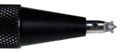 ACCUTOME
INTRODUCES NEW LIMBAL RELAXING INCISION KNIFE.
ACCUTOME
INTRODUCES NEW LIMBAL RELAXING INCISION KNIFE.
|
||||||||||||||


|
||
|
||||||||||||||


| Subscriptions: Review of Ophthalmology Online
is provided free of charge as a service of Jobson Publishing, LLC. If
you enjoy reading Review of Ophthalmology Online, please tell a
friend or colleague about it. Forward this newsletter or send this address:
[email protected].
To change your subscription, reply to this message and give us your old
address and your new address; type "Change of Address" in the
subject line. If you do not want to receive Review of Ophthalmology
Online, reply to this message and type "Unsubscribe: Review of Ophthalmology
Online" in the subject line. Advertising: For information on advertising in this e-mail newsletter or other creative advertising opportunities with Review of Ophthalmology, please contact publisher Rick Bay, or sales managers James Henne, Michele Barrett or Kimberly McCarthy. News: To submit news, send an e-mail, or FAX your news to 610.492.1049 |


