Since the introduction of accommodative intraocular lenses, much of what we know about how these lenses work has been based in theory. It has never really been possible to witness an accommodative IOL in vivo as an eye changes focus from distant to near. This is no longer the case.
For the past several months, we have been working with an ultrasound system (Eye Cubed, Ellex) and have found that for the first time, we can actually visualize how accommodative IOLs such as the Crystalens (Eyeonics/Bausch & Lomb) function following implantation.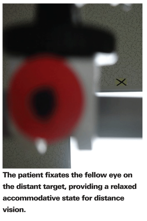
The difference between this ultrasound imaging system and others is its ability to capture 10-second, high-quality video images. And with a scan rate of 13 frames per second, it allows for real-time imaging of movement. With this video capture, we can see how the lens moves, and in some eyes, does not move. By performing this type of evaluation of the postoperative movements of the Crystalens, we hope to develop criteria that will tell us in advance if a patient may not be an ideal candidate.
The Premise of Crystalens
The concept of the Crystalens is that it provides pseudophakic accommodation by moving during ciliary body contraction, which causes an increase in vitreous pressure that triggers the IOL to move anteriorly.
However, we have never really had the ability to directly assess if the CrystaLens, or any accommodative IOL, is providing true pseudophakic accommodation or pseudoaccommodation. We could measure accommodative shift using wavefront analysis, while a slit-lamp examination could detect microscopic movements. Both traditional ultrasound imaging and optical coherence tomography systems are capable of capturing only static images.
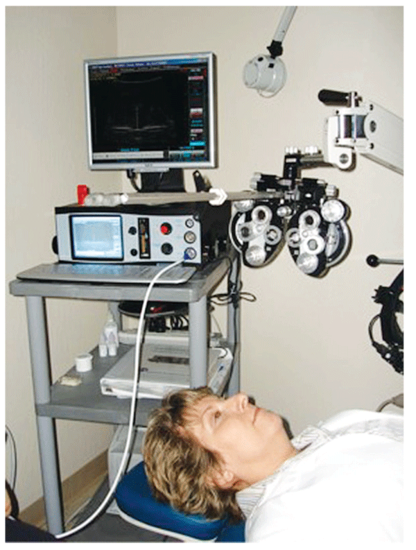
Figure 1. Dr. Rivera"s "CrystaLens Lab."
The Eye Cubed is capable of high-resolution video ultrasonography, which provides the missing piece when evaluating the movement of an accommodative IOL—a 10-second video clip of movement in the anterior segment. With this video capture we are now able to see the movement of the lens, as well as some movement of the iris as it comes forward and away from the capsular bag during the accommodative process. You can take that 10-second clip and go frame by frame to do an analysis with metrics and thereby determine the degree of movement, the amount of anterior displacement of the lens. For the first time we are developing a greater understanding for how this lens works in the eye.
The Crystalens Lab
To better understand how the accommodating process works, as well as to gain some insight into why the Crystalens does not work in every eye, we've developed what I call the Crystalens Lab. Our lab consists of the Eye Cubed System along with near and far point fixation targets (See Figure 1). To evaluate the IOL, we position the patient in a supine position with the subject eye under immersion ultrasound with the Eye Cubed. The patient fixates the fellow eye on the distant target. This provides us with a relaxed accommodative state for distance vision. We then have the patient fixate on the near target and perform a video capture. The set-up works extremely well, and patients tolerate the procedure without any discomfort.
Troubleshooting
The whole premise of the Crystalens is that it must move in order to provide pseudophakic accommodation. Yet, in some patients implanted with this IOL, the lens simply does not move. With the Eye Cubed, we've been able to observe these lenses and then successfully treat the problem that was hindering movement. Among them:
• Elschnig's pearls. We've identified things as simple as Elschnig's pearls postoperatively. If there is a lot of capsular debris at the end of the case that can form Elschnig's pearls later on, we've been able to see that those pearls can infringe on the ability of the lens to move. We've been able to see in those patients with pearls that the lens just sits there and they don't achieve pseudophakic accommodation (See Figure 3).
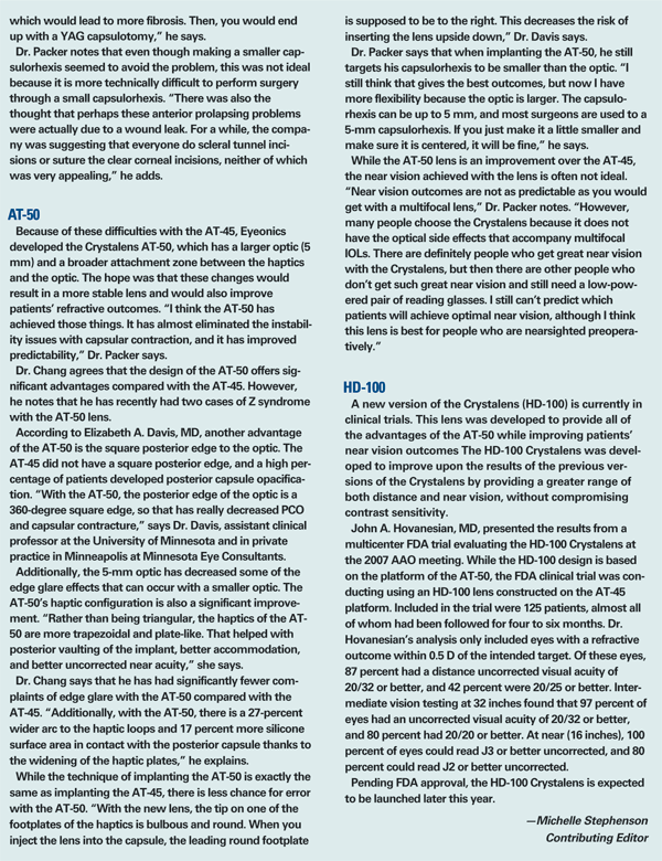
• Vitreous strands. One of the first cases where we used the Crystalens lab was on a patient who had undergone Crystalens implantation and then went to develop posterior capsular opacification. She underwent an Nd:YAG laser capsulotomy after which vitreous came forward, causing pupillary block and requiring a laser iridotomy. She wasn't happy with her accommodation afterwards. 
With the Eye Cubed, we were able to clearly see that the vitreous had come forward and essentially had encased the lens to the point where its movement was being restricted (See Figure 4). She also had a strand of vitreous that was free, so with accommodation, you could see this strand pulsing forward in front of the lens (See Figure 4). Before, we just could not have visualized that. She's since undergone a vitrectomy and is doing quite well, with a return of accommodative function of the lens.
• Ciliary insufficiency. This is a new phrase that we've adopted to describe eyes in which the patient has no accommodative effect with the Crystalens. In the small number of patients in whom we've encountered this, it would appear that the ciliary muscles are not as well developed. In one patient where Eye Cubed imaging was used, it was quite obvious that the ciliary muscle was underdeveloped (See Figure 5). If we are able to show that this entity actually occurs in a larger group of patients, we might be able to screen ahead of surgery and know whether the accommodative lens will be an effective choice or not.
Textbook Cataract Surgery
If there is one point that has become quite clear to me during our initial work with the Eye Cubed and the Crystalens, it is that this IOL is very particular when it comes to the status of the capsular bag at the end of the case—It must be in pristine condition. 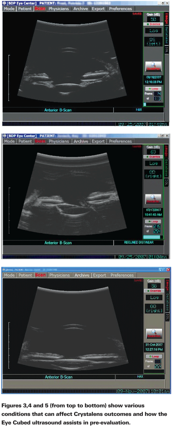
I now make very certain to do a very thorough I/A cortical cleanup at the end of each case. I also rotate the IOL once it is placed inside the bag to make certain that there are no loose fragments out in the periphery. There must not even a small tear in the capsulotomy. In one patient, a small, radial tear at the end of the case resulted in the Crystalens coming out of the bag and into the sulcus, leaving the patient with no accommodative effect. This was clearly visualized with the Eye Cubed.
It is exciting to now have a tool that allows us to more fully evaluate the accommodative effect. The use of this technology will certainly lead to greater predictability and effectiveness with this IOL.
Dr. Rivera is a partner in the


