


Rosacea patients often confrontthe ophthalmologist with secondary ocular symptoms consisting of burning, itching, foreign body sensation, conjunctival hyperemia and photosensitivity. The ocular findings are primarily confined to the eyelid or to the ocular surface1 causing conjunctivitis and eyelid irregularities.2
The majority of patients have concomitant cutaneous findings, which primarily affect the convexities of the central face (cheeks, chin, nose and central forehead) with flushing, erythema, telangiectasia, edema, papules, pustules, ocular lesions and rhinophyma.3
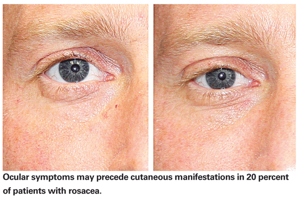
Ocular symptoms, however, may precede cutaneous manifestations in 20 percent of pa-tients with rosacea. Often times, the recalcitrant cutaneous telangiectasias do not respond well to topical or oral treatments and patients desire alternative treatment options.
The precise etiology of rosacea remains unknown,3,4 however, the disease is characterized by remissions and exacerbations of inflammation primarily effecting vascularity of sun-exposed sites on facial skin in genetically predisposed individuals.
The vascular dilation causes increased blood flow with resultant telengiectasia of the skin and interpalpebral area.
Ocular rosacea treatment is directed at protecting the surface with lubrication and minimizing inflammation to the ocular surface and eyelid region. Patients may present to the ophthalmologist first for their ocular symptoms and knowing how to effectively eradicate their cutaneous telangiectasia with laser therapy can be beneficial both for the patient and the physician.
Cutaneous Rosacea
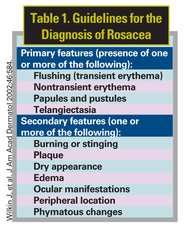
Skin manifestations of rosacea typically occur in patients in their third decade of life. The primary features observed with cutaneous rosacea are erythema, edema, papules, pustules and telengiectasia (See Table 1). Skin manifestations in patients can occur in different configuration that often change over time as flare-ups interrupt periods of remission.5
Typically, rosacea patients are followed and treated by dermatologists. The initial treatment modality incorporates topical treatments such as 0.75% metronidazole (Metrogel) or 1% metronidazole (Noritate cream).
Patients with pustular lesions are treated with sulfacetamide sulfur lotions and oral doxycycline may be added for recalcitrant cases. The efficacy of antibiotics has been substantiated, however, the chronic nature of rosacea makes long-term use undesirable.
Recent studies suggest that a low, subantimicrobial dose of doxycycline may provide efficacious treatment with a low side effect profile.6
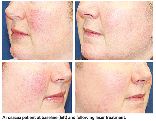
Often patients will have prominent telangiectasia, which do not respond to either topical medication or oral antibiotics. These are the patients that are referred to cosmetic surgeons for laser treatment.
Light-Based Treatments for Rosacea
When the effects from the topical medications have plateaued then patients can seek laser or light-based treatments.
The erythematous component of rosacea can be effectively addressed with intense pulsed light. This broadband light source targets the small vessels involved with the background erythema. A pulse dye laser (585 to 595 nm) with a small pulse duration also works well to clear the vessels.
The larger telangiectasias are most effectively treated with the pulsed dye laser, the first laser designed using the theory of selective photothermolysis for the treatment of vascular lesions. This theory states that a selected wavelength is chosen for treatment based on its ability to maximize optical absorption and heating in the target chromophore.
It further states that the energy delivered must be high enough to destroy the target and delivered with a pulse duration that is less than the thermal relaxation time of the target.
This ensures that the adjacent tissue surrounding the target is spared.7
Optical scattering should also be considered when formulating treatment because the depth to which incident radiation penetrates tissue is determined by a combination of both optical absorption as it relates to wavelength and optical scattering, which can be effected by spot size.8,9 We typically use a 7-mm spot size when delivering pulsed dye and Nd:YAG laser.
So why has the pulse dye laser been the gold standard for the treatment of vascular lesions?
The target chromophore for treating telangiectasia is hemoglobin. The pulse dye laser has a wavelength that is the closest to the absorption peaks of hemoglobin at 541 and 577 nm.
Prior to the development of the pulsed dye laser, the argon laser was used to treat blood vessels. The argon laser has blue-green light with a 514-nm wavelength which is absorbed by the hemoglobin molecule in red blood cells. Ophthalmologists still use this laser to treat retinal neovascularization.
The argon showed limited success with the treatment of cutaneous blood vessels. The larger blood vessels of the face responded moderately well, but the blood vessels contained within port wine stains differed physically and optically from the facial telengiectasia and therefore were unresponsive. Because of the shorter wavelength of the Argon laser, only superficial vessels could be treated and often times the epidermal skin would be victimized.
The long wavelength pulse dye laser (595 nm) bypasses the epidermal melanin and targets the hemoglobin in blood vessels. Lower melanin absorption also decreases energy loss in the epidermis, which improves the overall efficiency.10
For vessels situated deeper in the dermis, the wavelength of choice would be the Nd:YAG laser. Hemoglobin has strong absorption peaks at 541 nm and 577 nm and has secondary peaks at 800 nm and 1,000 nm. Although the pulse dye laser has a stronger absorption coefficient over the Nd:YAG, the longer wavelength of the Nd:YAG allows for greater penetration. Clinical efficacy in the clearance of vessels by the Nd:YAG can be demonstrated with higher fluences.
Vessels with greater diameter located deeper in the tissue plane are hard to treat with pulse dye laser. The Nd:YAG laser can penetrate deeper, however higher fluences are needed, which can contribute to patient discomfort.
When hemoglobin is exposed to the pulse dye laser, it is converted into methemoglobin (met-Hb). The Nd:YAG laser has a three-to-five times greater absorption of met-Hb than oxyhemoglobin,11,12 therefore lesser fluences are needed to achieve the threshold of coagulation, which correlates to greater efficiency in vessel clearance along with greater patient comfort.
The technology that I prefer incorporates these two wavelengths in a simultaneous or multiplexed fashion (Cynergy Laser, Cynosure,
This modality allows for better clearance of the recalcitrant telangiectatic vessels of rosacea over pulsed dye laser alone.13
Treatment Parameters
For patients with prominent erythema and telangiectasia, we typically treat the erythema first with intense pulsed light (Lumenis One, Lumenis,
Cold air is also blown over the face to further protect the epithelium. Longer pulse durations are selected to allow for gentle heating of the erythematous portion. Because broad band light contains non-coherent light from 530 to 1,200nm, the small vessels contributing to the flushed erythematous appearance are targeted.
Larger blood vessels are then treated with a combination laser that employs pulsed dye laser and Nd:YAG (Cynergy). Cold air is incorporated into the hand piece and the fluence is selected for both the PDL and Nd:YAG. The vessel is traced with the laser pulses beginning at the distal end of the blood flow to shut down the offending vessel.
Pulses are aligned in an interlocking fashion with minimal overlap. As the vessel is treated, the end-point is the appearance of cyanosis or bluish coloration change to the vessel. This indicates that an appropriate amount of wavelength specific energy has been absorbed by the target to create a small clot. Often times, the smaller vessels will rupture and mild purpura will be noted.
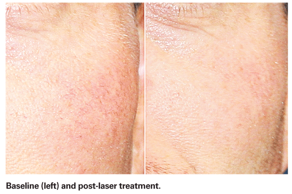
Typically one to three treatments are necessary to clear the vessels. Patients are followed one month post-laser to check for clearance. If vessels persist, then an additional treatment is performed. Patients are advised to wear sunscreen post-treatment for at least two weeks.
Since rosacea is a chronic disease, patients are also advised that erythema and telangiectasia can reoccur in the future. Continuing on their medical regimen prescribed by their dermatologist is necessary.
Dr. Saluja, a cosmetic surgeon and board-certified ophthalmologist, completed her cosmetic surgery fellowship through the
1. Browning DJ, Proia AD. Ocular rosacea. Surv Ophthalmol 1986;31(3):145-58.
2. Macsai MS, Mannis MJ, Huntley AC. Acne rosacea. In: Eye and skin disease.
3. Wilkin JK. Rosacea: Pathophysiology and treatment. Arch Dermatol 1994;130:359-62.
4. Tisma VS, Basta-Juzbasic A, Jaganjac M, et al. Oxidative stress and ferritin expression in the skin of patients with rosacea. J Am Acad Dermatol 2009;60:270-6.
6. Bikowski JB. Subantimicrobial dose doxycycline for acne and rosacea. Skinmed. 2003 Jul-Aug;2(4): 234-45. Review.
7.
8.
9.
10. Ross E, Domankevitz Y. Laser Treatment of Leg Veins: Physical Mechanisms and Theoretical Considerations. Lasers in Surgery and Medicine 2005;36:105–116.
11. Barton JK, et al. Optical and magnetic resonance changes in photothermally coagulating blood. Proceedings of SPIE 2002; 4609:10-19.
12. Kuenstner JT, Norris KH. Spectrophotometry of human hemoglobin in the near infrared region from 1000 to 2500 nm. Near Infrared Spectrosc 1994;2:59-65.
13. Karsai S, Roos S, Raulin C. Treatment of Facial Telangiectasia Using a Dual Wavelength Laser System (595 and 1,064 nm) A Randomized Controlled Trial with Blinded Response Evaluation. Dermatol Surg 2008 May;34(5): 702-8. Epub 2008 Mar 3.


