Though LASIK is an extremely effective and safe procedure for the right patient, there's always the possibility of a complication. Epithelial cells can begin to migrate beneath the flap, flap striae can develop or your femtosecond laser can produce bubbles in the anterior chamber that block your excimer laser's tracker. Here's how you can avoid and manage some common mishaps.
Ingrowth and Debris
If you notice epithelial cells beneath the flap, the first thing to do is identify their source and determine if they're coming from the flap edge. Instead of emanating from the edge, they may just be implanted epithelial cells from the surgery itself. These implanted cells are usually innocuous and will die off by themselves. However, you may encounter ingrowth coming from the edge of the flap where the cells are proliferating. At the edge, they create a fistula through which they grow beneath the flap, migrating toward the pupil. Note that an epithelial defect during surgery increases the risk of fistula formation. Also, the risk of epithelial ingrowth is higher after LASIK enhancement than it is for the initial LASIK procedure, because the edge of the flap isn't as neatly cut as it is for the first procedure.
Fluorescein staining may reveal a tiny, pinpoint stain at the edge of the flap, indicating that there's still an active fistula. If you see this, follow the patient more closely than you would someone without a fistula. Watch the patient to see where the ingrowth is going, and always assume that the ingrowth is more advanced than what you see at the slit lamp: There may be a sheet of epithelial cells that is a leading edge beyond the edge that you can actually see. However, as long as the vision isn't affected and the cells are away from the visual axis, I usually won't intervene since many of these cases will regress on their own. If the ingrowth doesn't resolve, however, I'll lift the flap and scrape the cells off of both the stromal bed and the underside of the flap using a spatula or dry micro sponges.

You may also encounter debris beneath the flap, which you can avoid by irrigating copiously before lifting. If we see any debris at the slit lamp for the 30-minute postop exam, we'll irrigate them out then and there. On many occasions, we'll see meibomian gland secretions under the flap, but those tend to be harmless.
Flap Striae
When I notice striae in the flap, I first need to determine if they're visually significant or not. If I can refract the patient to a crisp 20/20, then I know the striae probably aren't visually significant. Sometimes, mild striae will weakly affect the vision but will get better with time as the epithelium grows over them and fills in the small folds. However, if the patient is still complaining despite the refraction, and the vision isn't correctable, those are signs that I need to intervene, and the sooner the better.
The first stage of intervention is to lift the flap, preferably on day one, and hydrate it with saline solution. When the solution swells the flap, it can stretch and eliminate the striae by itself. You can also use a spatula to iron the flap and try to flatten the folds that way.
If the first stage doesn't work, you may need to remove the epithelium over the striae, because it may have grown into the small crevices of the striae and, as a result, will make stretching difficult. Using alcohol to loosen the epithelium before scraping it off is one effective strategy.
If the second stage is unsuccessful, the next step is to place sutures. I usually place seven diametrically opposed sutures at the flap edge—not the hinge—and leave them there for about three weeks. When you place them, they can go in at about 30 percent of the corneal thickness. They shouldn't be very tight, either, to avoid inducing astigmatism.
Femtosecond Foibles
If you use a femtosecond laser to create the flap, here are some problems you may encounter. I base my advice on experiences with the IntraLase.
• Opaque bubble layer.This layer of bubbles in the flap interface occurs on occasion, and is unavoidable. The OBL interferes with the ability of intraoperative pachymetry to accurately measure the stromal bed, and with the ability of the excimer's eye tracker to work properly. The latter is more of an issue with lasers that don't allow you to deactivate the tracker, such as the Alcon Allegretto.
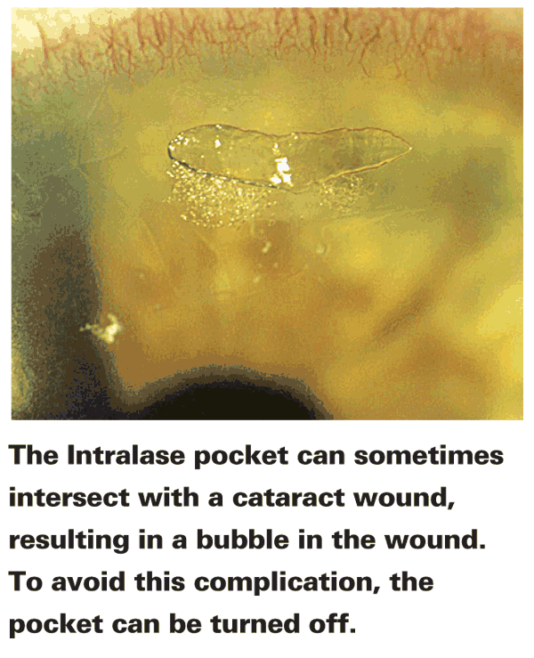
There are certain things that make OBL more frequent. One such factor is turning off the pocket creation function of the laser. The pocket is a small space created in the cornea to receive the bubbles during flap creation so an OBL doesn't form. In some cases, you may want or need to turn off the pocket in order to increase the diameter of the flap. Doing so increases your risk for an OBL.
• Anterior chamber bubbles. Bubbles generated during flap creation can trickle through the trabecular meshwork and enter the anterior chamber, also affecting the excimer's tracking mechanism. If these occur and you have a laser with which you can't deactivate the tracker, you may have to have the patient wait for two hours while the bubbles are resorbed.
• Diffuse lamellar keratitis. DLK can occur if your energy levels are too high, so you may have to adjust them to reduce the risk of DLK. One thing we've found is that if you put the flap orientation ink marks on the side of the flap into which you're going to enter it to lift it, you can drag this ink into the interface and increase your risk of DLK. For that reason, I place my ink marks on the opposite side.
Obviously, treatment of DLK involves early detection and steroids. In cases of aggressive DLK, I'll use a Medrol dose pack of oral steroids.
If there are any signs of coalescing inflammatory cells in a case of DLK, I won't hesitate to lift the flap and irrigate beneath it. If I don't, the cells can lead to stage 4 DLK: melting of the cornea and scarring.
A new consideration with the Intra-Lase accompanies the use of very thin flaps. With a thin flap, you may be fooled by DLK that appears to be very superficial—too superficial to be DLK—and you have to keep that possibility in mind when you make your diagnosis. Recently, we had a case where the inflammatory cells seemed too close to the epithelium to be DLK. When we looked at the flap thickness, however, it turned out to be 70 µm. That explained why the cells were so superficial, and prompted us to go back and irrigate them out.
• Decentered flap. To avoid decentered flaps, I look at the eye under the excimer microscope first, and put a dot of ink at the pupil center using a Sinskey hook. Then I decenter the suction superiorly and nasally. As I'm docking, I make sure that I'm always centered going down with the docking cone. As I'm going down with the cone, I make sure I'm centered on the ring of light reflected on the cornea rather than the pupil.
Once the docking occurs, I make sure that I don't have asymmetric pressure on the suction cup as the docking cone is pushing down on the cornea, which helps prevent decentration. Anytime I feel asymmetric pressure, I readjust.
Once docked, I'll compare the location of the ink dot to the reticule of the laser. I don't necessarily need to align the central reticule to the dot itself, because if you move it too much it will decrease the diameter of the flap. Instead, I move it enough just so that it's in the vicinity of the dot. I don't use the pupil as a guide, because it's distorted by the applanation and therefore isn't a good guide for centration.
If I'm too decentered and have to move the flap significantly, it may decrease the size of the flap. If the flap is smaller than 8.8 mm, I'll turn off the pocket. This gives me an additional 0.1 to 0.2 mm diameter but increases the risk of OBL. I start with a flap of 9.3 mm, which gives me more leeway in terms of movement of the reticule.
Even the best surgeons run into problems. I hope this advice helps you deal with those times when complications sully your surgical day.
Dr. Melki is an attending physician at the


