Presentation
A 57-year-old white male with no past ocular history presented to the Wills Eye Emergency Room with difficulty focusing, "jumping" eye movements and double vision for 24 hours. The double vision was worse in right gaze and improved with left gaze. It improved with covering either eye. The images were displaced diagonally. There was no noticeable difference in symptoms at distance versus near. The patient stated that his symptoms began suddenly during a meeting at work. His symptoms had been constant and non-progressive since that time. He denied a change in visual acuity or eye pain, but did admit to a generalized difficulty in focusing. He denied a history of trauma.
Medical History
The patient had a past medical history of hyperlipidemia, gastroesophageal reflux disease, genital herpes and anxiety. He denied a past ocular history. He did not have any history of diabetes, hypertension, renal failure or hypercoaguability. His medications included ezetimibe/simvastatin, valacyclovir and lorazepam. He was not a smoker. His family history was noncontributory. He denied a history of previous or current vision loss, eye pain, headache, weakness, parasthesias, facial droop, slurred speech or ataxia.

Examination
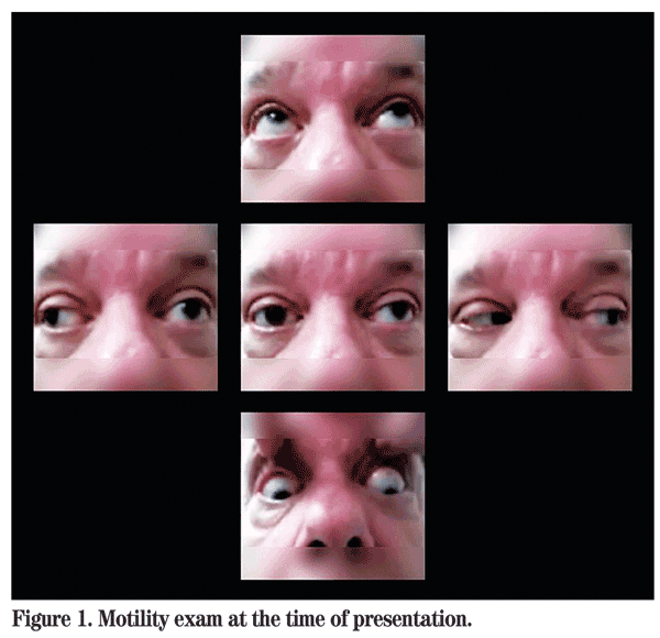
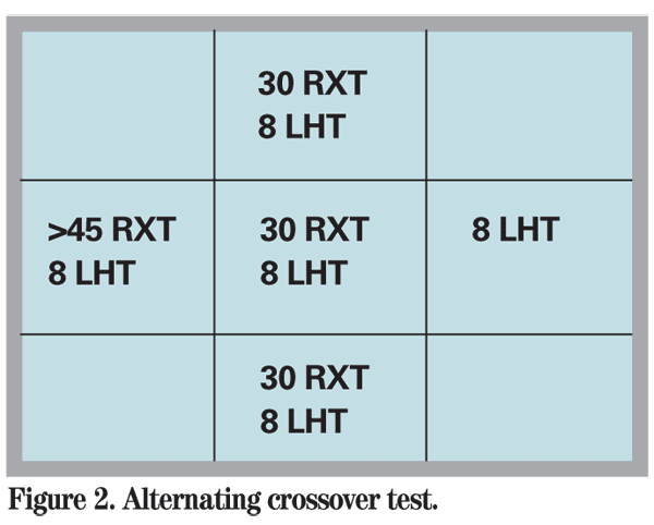
The patient's examination at the time of presentation revealed a near uncorrected visual acuity of 20/70 in the right eye and 20/100 in the left eye. There was no relative afferent pupillary defect or anisocoria. Visual fields were full to confrontation. No ptosis was noted. Motility photos were obtained at the time of initial presentation and are shown below (See Figure 1). Alternating cross-cover test was preformed and demonstrated a comitant left hypertropia and incomitant exotropia (worse in right gaze) (See Figure 2). The patient was noted to have nystagmus with both vertical and torsional components: Intorsion was observed with elevation; and extorsion with depression. Abducting nystagmus was evident in the right eye on right gaze. Intraocular pressure was 16 mmHg in both eyes. Slit-lamp exam and fundus exam were within normal limits in both eyes. Neurologic exam did not demonstrate any other focal deficit.

Diagnosis, Workup and Treatment
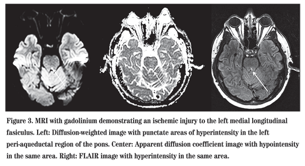
The patient presented with acute onset of left internuclear ophthalmoplegia (INO), see-saw nystagmus and skew deviation. This localizes the lesion to the brainstem, particularly to the midbrain and left medial longitudinal fasiculus (MLF). Differential diagnosis for this lesion includes a demyelinating disease, such as multiple sclerosis, brainstem stroke, trauma, metastatic lesion or primary tumor. Despite oral sedation, the patient was unable to tolerate a standard MRI because of claustrophobia.

Discussion
In anatomic terms, an internuclear lesion refers to a lesion between two cranial nuclei. Internuclear ophthalmoplegia denotes a lesion in the medial longitudinal fasiculus, a bundle of fibers that connect the sixth nerve nucleus of the pons to the contralateral medial rectus subnucleus of the midbrain.
The primary feature of INO is limited or slowed adduction of the ipsilateral eye. The contralateral eye often shows abducting nystagmus, which our patient demonstrated. A skew deviation can also be caused by a lesion in the MLF, and therefore may accompany an INO. Our patient had a left hypertropia consistent with a skew deviation. Lastly, our patient had "see-saw" nystagmus. This is a rare nystagmus characterized by simultaneous elevation and intorsion of one eye, and depression and extorsion of the other. It localizes to the parasellar region or midbrain. Therefore, all three features of his exam localize to the left brainstem, and particularly midbrain, consistent with the location of his infarct at the left pontomedullary junction.
Differential diagnosis is as mentioned, with demyelinating lesions being the most common in younger patients and ischemic lesions being the most common in older patients. INO may be the only sign of acute brainstem stroke. Myasthenia gravis may cause a pseudo-INO or pseudo-one-and-a-half syndrome.
Older patients with an isolated acute INO and no history of MS symptoms deserve rapid MRI neuroimaging to evaluate for stroke. Recovery of binocular fusion is expected in most patients with ischemic INO. Those with other neurologic symptoms generally take longer to recover.
The author would like to thank Jennifer Hall, MD, of the Wills Eye Neuro-Ophthalmology Service for her time and assistance with this case, and Joshua Smith for his assistance with image processing.
1.
2. Kanski JJ featuring Burton B. Supranuclear disorders of ocular motility. In: Kanski JJ, ed. Clinical Ophthalmology, A Systematic Approach: Chapter 21, Neuro-ophthalmology. Elsevier; 2007:825-6.
3. Deleu D, Sokrab T, Salim K, El Siddig A, Hamad AA. Pure isolated unilateral internuclear ophthalmoplegia from ischemic origin: Report of a case and literature review. Acta Neurol Belg 2005;105:214-7.
4. Kim JS. Internuclear ophthalmoplegia as an isolated or predominant symptom of brainstem infarction. Neurology 2004;62:1491-6.


