


 Q: I have a 51-year-old black female patient with a recent onset of vertical diplopia that disappears when either eye is closed. Where do I go from here?
A: First, determine the cause of the diplopia. Since it disappears when either eye is closed, we can conclude that there is ocular misalignment, says Patricia A. Modica, O.D., assistant clinical professor at SUNY State College of Optometry. Carefully observe eye movements in all diagnostic positions of gaze. Look for limitations that might suggest a muscle weakness.
The most common cause of vertical diplopia is superior oblique palsy. Look for a hypertropia that increases in contralateral gaze and ipsilateral head tilt. Also, observe the patient for a characteristic compensatory head tilt, Dr. Modica says.
Distinguish this from other origins, such as:
Third nerve palsy, which likely has both a vertical and a horizontal component.
Skew deviation, which usually appears with concomitant hypertropia and other abnormal eye movements, such as nystagmus.
Diseases of the orbit, which include neoplastic, inflammatory, infectious or traumatic processes. Proptosis, lid retraction, periorbital edema, conjunctival hyperemia and sometimes disc edema should tip you off to this process, Dr. Modica says.
Finally, consider myasthenia gravis (MG) in any diplopia case, she says. Myasthenia gravis is an autoimmune disorder in which antibodies prevent the neurotransmitter acetylcholine from attaching to muscle receptors, thereby interfering with muscle contractions. The disorder is characterized by fatigability of voluntary eye movements. Patients typically complain of diplopia that worsens as the day progresses, and improves after sleep, she says.
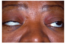
Ptosis and ocular misalignment can be clues to myasthenia gravis.
The ocular form of MG is limited to the ocular muscles and can present with diplopia, ptosis, or both. MG may even be a possibility when the patients motility presentation is identical to that seen in any extraocular muscle palsy, so a careful case history is crucial to make the correct diagnosis.
If you suspect MG, Dr. Modica says, try a few simple in-office tests:
Sustained upgaze test. On sustained upgaze, patients who present with myasthenic ptosis can show gradual worsening as the levator muscle fatigues.
Ice pack test. Apply a cold pack to the eyelid for five minutes and check for improvement of ptosis. Cold temperatures allow acetylcholine to have more time to react with the muscle receptor sites to enhance muscle contraction and function, Dr. Modica says.
Sleep test. Since myasthenics typically improve after sleep or rest, simply have the patient take a nap in the exam chair for 30 to 45 minutes, she says.
More formal diagnostic procedures include serologic and electrodiagnostic studies. However, most clinicians still rely heavily on the Tensilon test to make the diagnosis of ocular MG. Tensilon (edrophonium, Valeant Pharmaceuticals) is an intravenous, cholinergic drug that typically shows improvement in muscle function within seconds.
Once diagnosed, treatment options for MG include the cholinergic drug Mestinon (pyridostigmine, Valeant Pharmaceuticals), Dr. Modica says. Immunosuppressive therapy is often necessary alone or in conjunction with Mestinon.
If medications dont effectively eliminate ptosis and diplopia, ocular management may consist of prismatic corrections and ptosis crutches, Dr. Modica says.
Comanage a patient who has ocular myasthenia with a neuro-ophthalmologist. If the myasthenia becomes generalized, the patient may be better served with a neurologist that specializes in neuromuscular disease, since management can be more tricky and complications (such as respiratory difficulty) can occur, Dr. Modica says.


